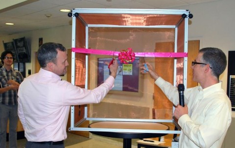

The Parker H. Petit Department of Bioengineering and Bioscience recently unveiled its newest core facility. But even before its official grand opening earlier this year, the Neuro Design Suite (NDS) was having an important impact on the work of researchers at the Georgia Institute of Technology.
Last year, when Craig Forest and Garrett Stanley applied for grant funding through the National Institutes of Health (NIH) for President Obama’s BRAIN Initiative (Brain Research through Advancing Innovative Neurotechnologies), they made sure to include the Neuro Design Suite in their description of available facilities.
It’s because facilities were among the important five criteria on which these grant proposals were scored by NIH, who awarded the Forest/Stanley research team $1.5 million as part of the first wave of BRAIN Initiative funding. Part of NIH’s reasoning on this score is that the NDS is a state-of-the-art facility with some of the best research tools available.
“They want to know, ‘does your team have adequate facilities to conduct this research?’ So, it made a great impression,” says Forest, associate professor of bioengineering in the George W. Woodruff School of Mechanical Engineering. “The modern tools of neuroscience are allowing researchers unprecedented access to the living brain at work. These tools allow measurements at the level of single cells, and the connections between them.”
Like all core facilities, the Neuro Design Suite are shared resources, a high-tech “sandbox” for engineers and scientists to try out their novel inventions in a controlled setting with all the necessary equipment at hand. So far, a number of different researchers from different disciplines have utilized the tools.
“Having a shared facility that can support multiple grants and multiple [principal investigators] is absolutely essential,” Forest says. “We’re excited that these tools invented for neuroscience could be brought to bear on entirely different problems.”
In other words, you don’t have to be a neuroscientist to reap the research benefits of the Neuro Design Suite. Assisting researchers who use the facility is lab manager Bo Yang, who can sit down with a scientist and help design experiments suitable to their needs or discipline.
“Bo has recorded the electrical activity of 2,500 brain cells,” says Forest. “We’re fortunate to have one of the world’s experts working with researchers.”
The suite features three major rigs that allow researchers to perform manual and/or automated in vitro, in vivo patch clamping and in vivo extracellular electrophysiology recordings.
The Q-Scientifica SliceScope within the in vitro patch clamping rig is a compact upright microscope equipped with a fully-motorized fixed stage, various electrode manipulators, a wide range of Olympus objectives and an LED system to meet the demands of electrophysiology study.
The electromagnetically shielded in vivo extracellular electrophysiology rig is constructed with various elements, including Zesis surgical microscopes, a 128-channel Tucker Davis Technologies data acquisition system (RZ2), Kopf stereotaxic frames, and DC temperature controllers to enable stable, reliable and high-quality recordings. Also, a complete LED driver system (Thorlabs) was equipped to this rig to facilitate optogenetic in vivo experiments.
Automatic patch clamping devices (autopatchers) are also attached to both in vitro and in vivo patch clamping rigs to obtain high yield and high quality whole cell recordings.
Forest and Stanley, professor in the Wallace H. Coulter Department of Biomedical Engineering (BME), were awarded BRAIN Initiative funding from the NIH for their project entitled, “In-vivo circuit activity measurement at single cell, sub-threshold resolution,” research that could only happen with the best tools available.
“We can use these tools not only to record what’s happening in cells, at the level of a single cell, but also in cells that are in two different brain regions simultaneously,” says Forest. “In each region we can record activity within a single cell, at the sub-threshold resolution of a single cell. No one’s been able to do that before. We can record cells talking to each other in a living brain.”
CONTACT:
Jerry Grillo
Communications Officer II
Parker H. Petit Institute for
Bioengineering and Bioscience
Jerry Grillo
Communications Officer II
Parker H. Petit Institute for
Bioengineering and Bioscience
